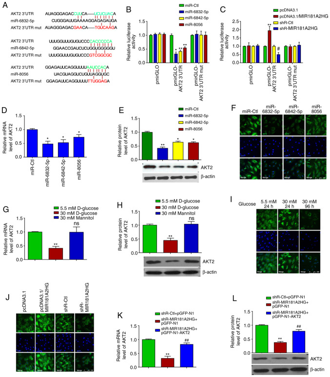
Figure 2.
miR-6832-5p, miR-6842-5p and miR-8056 target AKT2 3′UTR in HUVECs. (A) The predicted binding sites between miR-6832-5p, miR-6842-5p or miR-8056 and AKT2 3′UTR. Red color indicates the sequence of mutated sites. (B) Luciferase reporter assay was performed in HUVECs co-transfected with miRNAs, pRL-TK or pmirGLO-AKT2 3′ UTR (wild-type or mut). *P<0.05 and **P<0.01 vs. miR-Ctl. (C) Luciferase reporter assay was performed in HUVECs co-transfected with pmirGLO-AKT2 3′ UTR (wild-type or mut) and pcDNA3.1-MIR181A2HG or shR-MIR181A2HG. **P<0.01 vs. pcDNA3.1; #P<0.01 vs. shR-Ctl. (D-F) RT-qPCR, western blot analysis and immunofluorescence staining detected the relative expression of AKT2 mRNA and protein in HUVECs transfected with miR-6832-5p, miR-6842-5p, or miR-8056 mimics. *P<0.05 and **P<0.01 vs. miR-Ctl. (G-I) RT-qPCR, western blot analysis and immunofluorescence staining detected the relative expression of AKT2 mRNA and protein in HUVECs treated with 5.5 or 30 mM D-glucose for different time periods. **P<0.01 vs. 5.5 mM D-glucose. (J) Immunofluorescence staining detected the expression of AKT2 in MIR181A2HG-overexpressing or depleted HUVECs. (K and L) RT-qPCR and western blot analysis detected the expression of AKT2 mRNA and protein in MIR181A2HG-depleted or AKT2-overexpressing HUVECs. **P<0.01 vs. shR-Ctl+pGFP-N1; ##P<0.01 vs. shR-MIR181A2HG+pGFP-N1. HUVECs, human umbilical vein endothelial cells; ns, not significant.

