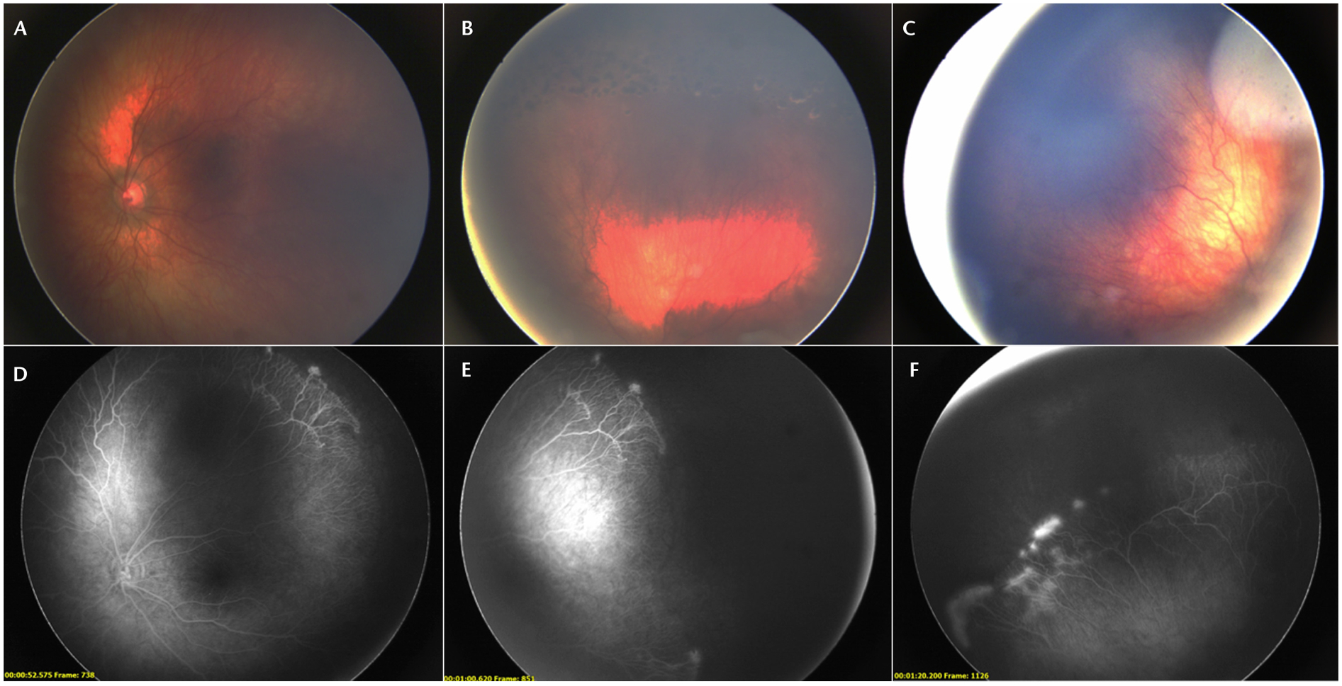
FIGURE 2.

Fundus photos (A to C) and fluorescein angiography (D to F) of a patient with retinopathy of prematurity who had previously been treated with laser. In the fluorescein angiography, signs of neovascularization and leakage are more obvious compared with the fundus photographs.
