
Fig. 2.
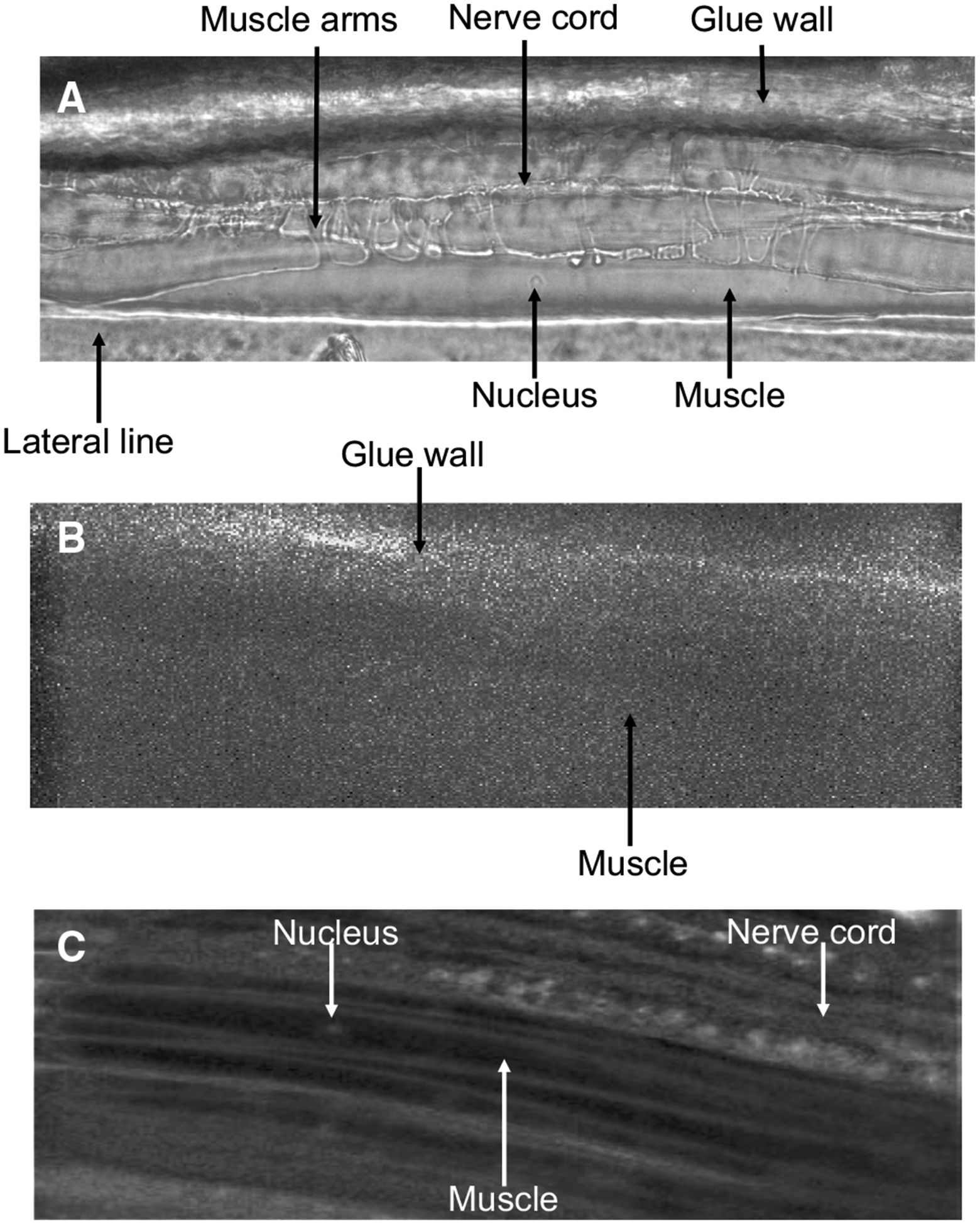
Micrographs of B. malayi muscles: a micrograph of B. malayi muscles under white light. b Untreated muscle exposed to blue light. c Muscles after being treated with 5 μM Fluo-3AM for 60 min at 34–36 °C. Key structures, muscle arms, muscle, nucleus, nerve cord, lateral line and glue wall are highlighted