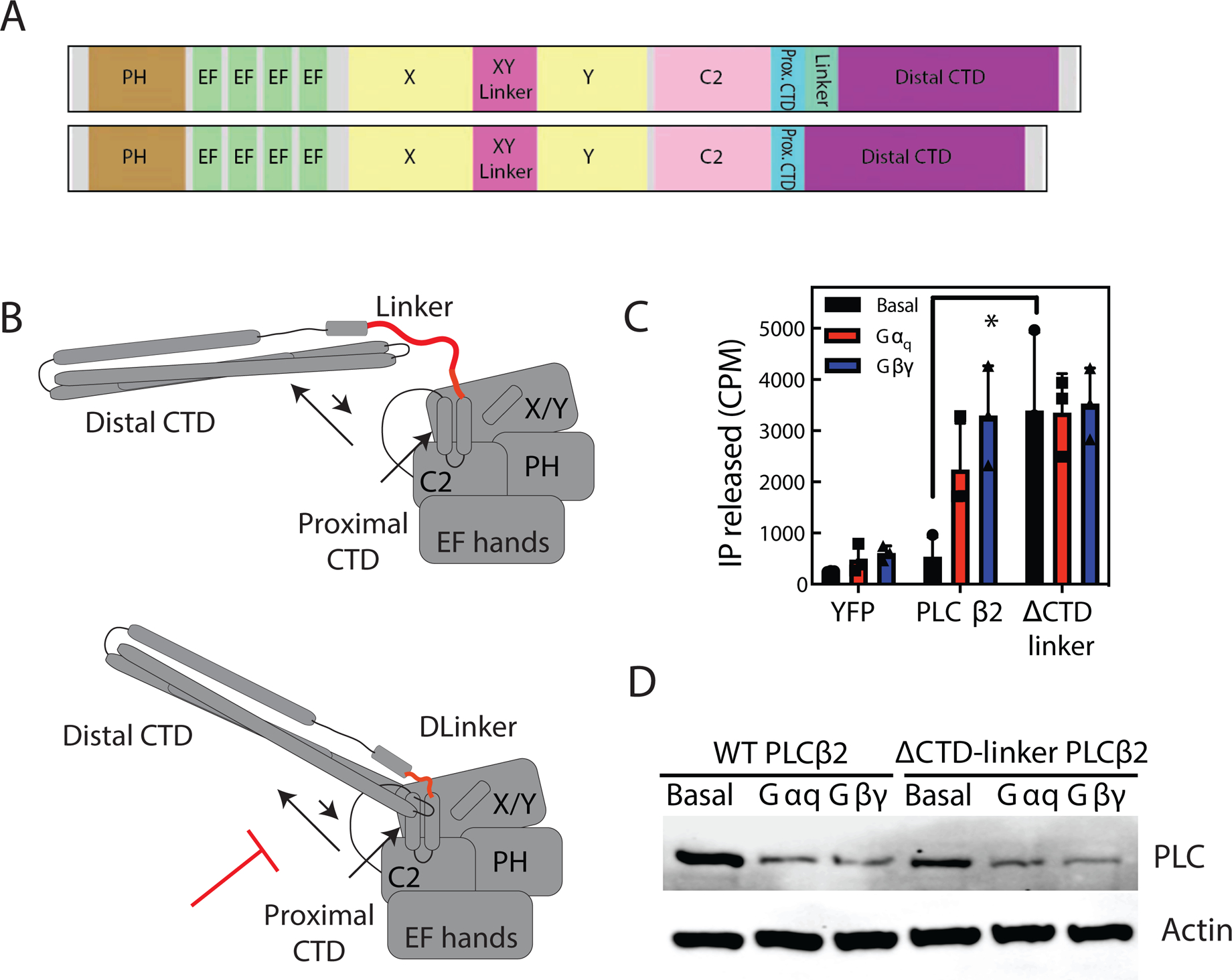
Figure 6: Restriction of Distal CTD movement increases PLC activity.

(A) Schematic of CTD-linker deletion (amino acids 839–872 deleted). (B) Cartoon diagram of model of CTD-linker deletion effects. By preventing C-terminal domain rearrangement, CTD linker would prevent autoinhibition. (C) Total inositol phosphate assay. COS-7 cells were transfected with 250 ng PLCβ2 WT or PLCβ2-ΔCTD-linker in the presence or absence of 200 ng Gβ1 and 200 ng Gγ2 or 200 ng Gαq, and total [3H] inositol phosphate accumulation was measured. The data shown are mean ± S.D. for at least three independent experiments and analysed by 2-way ANOVA with multiple comparisons. *, p < 0.05. (D) Western blot for PLCβ2 wt and PLCβ2-ΔCTD-linker mutant expressed in C, and actin as a loading control.
