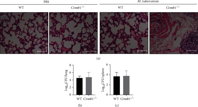
Figure 3.

Histopathology and bacterial load of lungs (and spleens) of mice with Mtb infection in vivo. (a) Representative histopathology in lung sections stained with haematoxylin and eosin (H&E) of wild-type mice and Ctnnb1−/− mice infected with or without M. tuberculosis for 4 weeks (bar, 100 μm; original magnification ×200). (b) Bacterial load (cfu) in the lungs of both Ctnnb1−/− (n = 5) and wild-type mice (n = 5) infected with M. tuberculosis for 4 weeks. (c) Bacterial load (cfu) in the spleens of both Ctnnb1−/− (n = 5) and wild-type mice (n = 5) infected with M. tuberculosis for 4 weeks. The experiment had three independent rounds, and results were analyzed by the unpaired Student t-test. PBS, phosphate-buffered saline.
