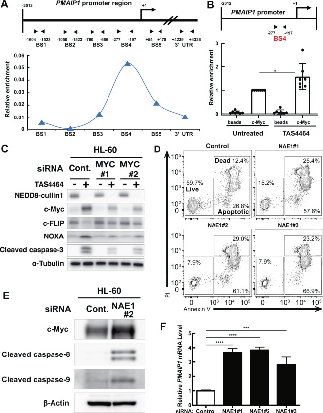
Fig. 6. TAS4464-induced c-Myc activation transcriptionally mediates apoptotic gene expression.
A Schematic representation of the PMAIP1 promoter region and c-Myc enrichment at each site. The regions targeted by the primer pairs are indicated as “BS1” to “BS5” and “3′ UTR”. B Enrichment of c-Myc protein in the BS4 region in (A). The fold enrichment value compared to the untreated condition was used to evaluate the effect of TAS4464 induction. HL-60 cells were treated with TAS4464 (0.1 μmol L−1) for 4 h. C MYC siRNAs were transfected into HL-60 cells. After 48 h, cells were treated with 0.1 μmol L−1 TAS4464 for 16 h except for c-Myc detection (1 h). Total protein was extracted and then immunoblotting for NEDD8-cullin1, c-Myc, c-FLIP, NOXA and cleaved caspase-3 was performed. D Apoptotic cell death was evaluated by flow cytometric analysis. HL-60 cells were treated with NAE1 siRNAs for 16 h. E Immunoblotting for c-Myc, Cleaved caspase-8 and Cleaved caspase-9 after NAE1 siRNAs transfection. Cells were harvested 8 h after transfection. F qRT-PCR to measure the level of PMAIP1 mRNA was performed in HL-60 cells after NAE1 siRNA transfection at 16 h time point. Data are presented as the mean ± SD values of data from three independent experiments. *P < 0.05, ***P < 0.001, ****P < 0.0001.

