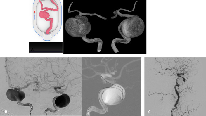
Figure 2.
(A) Cartridge (left) and surface rendered 3D-rotational angiogram (3DRA)(right) demonstrate 3D-printed model and working projects for navigation of right-sided giant cavernous IA. (B) DSA (left) and fluoroscopy roadmap image (right) demonstrate mimicked anteroposterior (AP) and lateral working projection views selected from the pre-procedural simulation that allowed for successful catheterization of the distal ICA. (C) DSA control run after coiling the aneurysm sac with jailed microcatheter, demonstrating total occlusion – Raymond–Roy scale I.20

