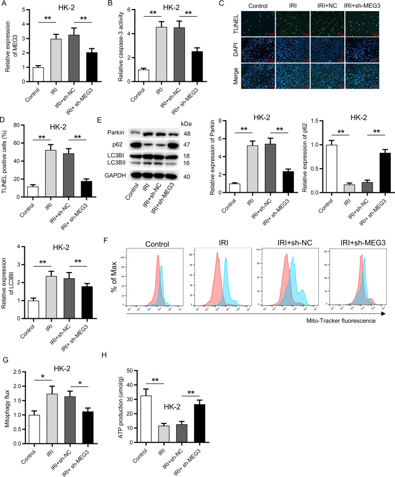
Fig. 2. Suppression of MEG3 inhibited apoptosis and mitophagy in vitro.
A RT-qPCR showed the transfection efficiency of sh-MEG3 in HK-2 cells. B A caspase-3 kit was applied to detect the activity of caspase-3 in sh-MEG3 transfected HK-2 cells. C, D A TUNEL assay revealed the influence of MEG3 deficiency on apoptosis of HK-2 cells. E Western blot analysis indicated the protein levels of Parkin, p62, LC3B-I, and LC3B-II. F, G MitoTracker Green FM staining was used to evaluate mitophagy flux under the influence of MEG3 deficiency. Red color indicated DMSO and blue color indicated Baf. H The influence of MEG3 deficiency on ATP production was determined via an XFe96 extracellular flux analyzer. *p < 0.05, **p < 0.01.

