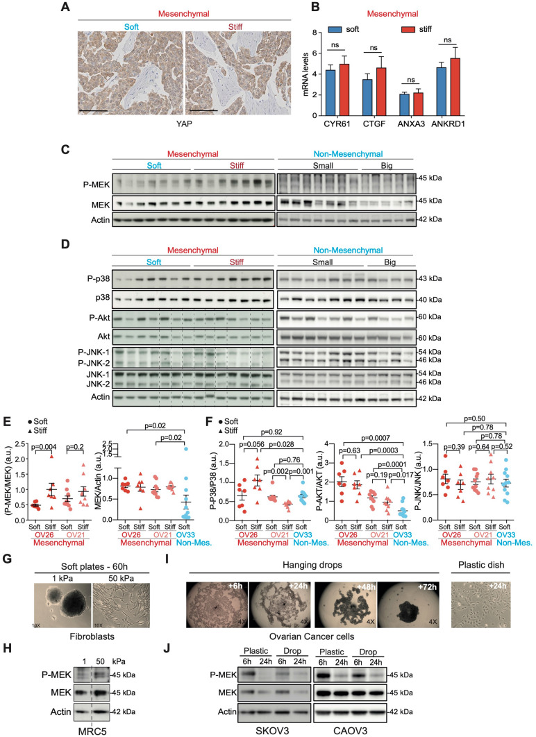
Figure 4.
MEK is activated upon tumor stiffening of mesenchymal HGSOC. (A) Representative views of YAP staining in soft (left) and stiff (right) Mesenchymal OV26 tumors. Scale bar: 100 μm. (B) Bar plots showing CYR61, CTGF, ANXA3 and ANKRD1 mRNA expression levels normalized to cyclophilin A in soft (n = 6) and stiff (n = 6) tumors. Data are shown as mean ± S.E.M. P values from Student’s t-test. (C) Representative western blots showing the phosphorylated form (P-) and the total protein levels of MEK in soft (n = 7) versus stiff (n = 7) Mesenchymal (OV26) and Non-Mesenchymal (OV33) (n = 12) HGSOC. (D) Same as in (C) for P38, AKT, JNK-1 and JNK-2 in soft (n = 7) versus stiff (n = 7) Mesenchymal OV26 and Non-Mesenchymal (OV33) (n = 11) tumors. Dashed lines are used to delineate different parts of two different gels run, blotted and revealed at the same time, with the same time of exposure. (E) Scatter plots of P-MEK/MEK and MEK/Actin ratios from soft (dot) and stiff (triangle) Mesenchymal (OV26: soft n = 7, stiff n = 7; OV21: soft n = 10, stiff n = 9) and Non-Mesenchymal (OV33, n = 12) tumors, as assessed by densitometry analysis of western blots shown in (C and Supplementary Fig. 2E). Data are shown as mean ± S.E.M. P values from Welch's t-test (left panel) and Mann–Whitney test (right panel). (F) Same as in (E) for P-P38/P38, P-AKT/AKT and P-JNK/JNK ratios (but n = 11 for OV33 Non-Mesenchymal tumors). (G) Representative images of human MRC5 fibroblasts, cultured 60 h either on soft (1 kPa—left) or stiff (50 kPa—right) polyacrylamide hydrogels. (H) Representative western blots showing P-MEK and MEK protein levels from MRC5 cells cultured as described in (G). Dashed line is used to delineate different parts from the same gel. (I) Representative images of ovarian cancer cells cultured in hanging drops for 6 h to 72 h or on plastic dish for 24 h. (J) Representative western blots showing P-MEK and MEK protein levels from SKOV3 and CAOV-3 ovarian cancer cell lines cultured 6 h or 24 h either on plastic plate (stiff) or in hanging drops (soft). Actin is used as an internal control for all protein loadings.

