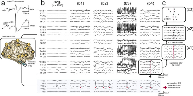
Figure 1.
Examples of scalp EEG waveforms associated with intracranial IEDs. (a) Scalp IEDs are less clear than their intracranial counterparts which results in inferior SNR. (b) Representative examples of incomplete discharge propagation (FCD + HS subject) in simultaneous scalp EEG and iEEG recordings. For clarity, only the right lateral temporal chain of scalp electrodes is illustrated. Automatically detected intracranial IEDs are marked by red dots under the intracranial portion of the signals (light blue background). Associated scalp waveforms are below (b1,b3) or near the limit of visual identifiability (b2,b4) but all were robustly detectable as a scalp zero-crossing pattern. (b2) shows trial-to-trial variability. (b3,b4) show how scalp EEG artifact or background rhythms interfere with visual detection. The first column (avg.) shows a small waveform resulting from signal averaging (c.f. Fig. 3a of Koessler et al.18). (c) Transformation of a multi-channel scalp EEG signal to a zero-crossing (z.c.) pattern (c3) through the steps of prefiltering (c1) and zero-crossing detection (c2).

