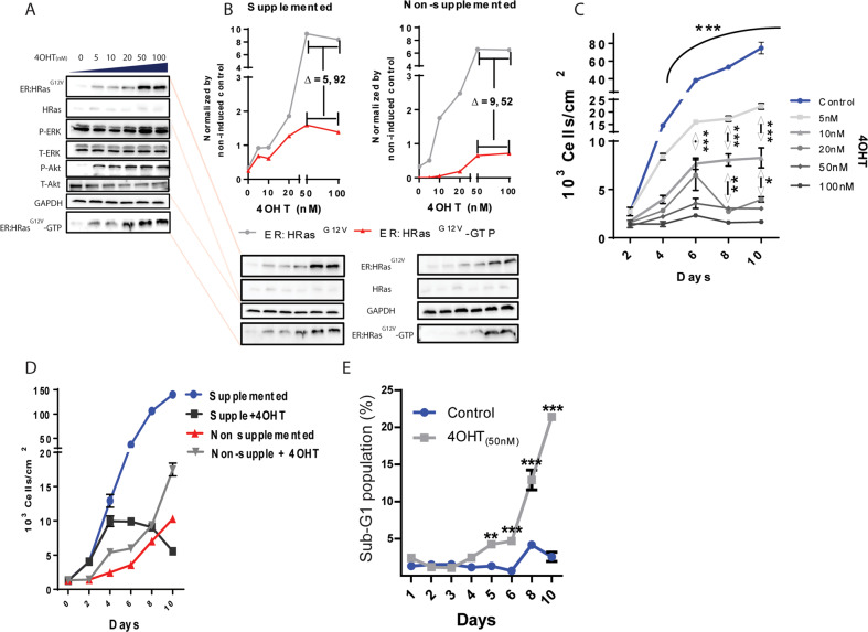
Fig. 1. High HRasG12V activity causes oncogenic stress in the E6E7 keratinocytes grown in supplemented media.
A Immunoblots: 4OHT dose–response of ER:HRasG12V induction. The activity of HRasG12V was monitored by a RAS-GTP-binding assay, which leads to downstream activation of P-ERK and P-Akt. B Immunoblots comparing ER:HRasG12V levels between supplemented (5 ng/mL of EGF and 5 µL/mL of pituitary extract) and nonsupplemented media in ER:HRasG12V-keratinocyte cultures. The immunoblot of cells in the supplemented medium is a projection of the one presented in A. The electrophoresis gel of cell lysates derived from cells grown in either supplemented or nonsupplemented media, was blotted in the same membrane to allow comparison of ER:HRasG12V levels and HRasG12V-GTP (HRasG12V-GTP = HRasG12V activity) under both conditions. C Growth curves of ER:HRasG12V keratinocytes in function of 4OHT concentration. The graph is representative of three independent experiments carried out in duplicate. D Effect of 50 nM 4OHT in ER:HRasG12V keratocytes growing in supplemented or nonsupplemented media. The graph is representative of three independent experiments carried out in duplicate. E Evolution of the sub-G1 population of ER:HRasG12V keratocytes growing in complete medium induced or not with 50 nM of 40HT, measured by flow cytometry after PI staining. The graph is representative of two independent experiments. For flow cytometry, 30 × 103 events were considered per condition and time point. Growth curves and the sub-G1 population are presenting data as mean (SD). Two-way ANOVA *P ≤ 0.05, **P ≤ 0.01, and ***P ≤ 0.001, Bonferroni post hoc.

