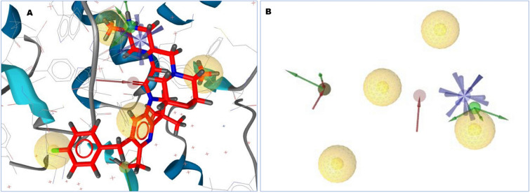
Figure 1.
(A) The 3D structure-based pharmacophore model of XIAP protein in complex with 46781908 (CID) ligands derived from the X-ray derived crystal structure of XIAP protein (PDB ID: 5OQW). (B) Several pharmacophore features were generated after complex interaction, four yellow spherical shapes indicating hydrophobic interaction, one blue star shape depicting the positive ionizable with tolerance 2, three red colors arrow and spherical shapes indicating H bond acceptor having tolerance 1.5, five hydrogen bond donors represented by green spherical or arrow shape have been identified within the protein–ligand complex interaction. 15 exclusion volume, which were generated during pharmacophore modeling has not showed in this schematic presentation.

