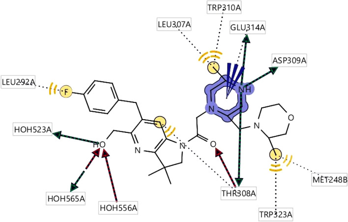
Figure 2.
2D structure obtained during the pharmacophore modeling showing the hydrophobic interaction depicting yellow color and the interaction with the amino acid residues in our selected XIAP protein. Hydrogen bond donor (HBD) features most frequently participated in ligand–protein interaction were shown in green color, whereas the red color showing the interaction of Hydrogen bond acceptors (HBA) to the oxygen, nitrogen atom of the benzine ring and its different side chains. Hydrogen atoms as well as restricted area maintain the shape and position of the binding pocket did not mention in the figure.

