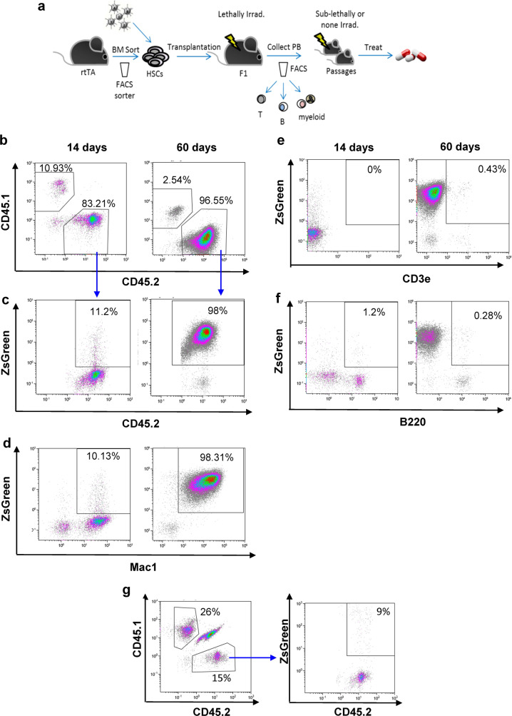
Fig. 1. Overexpression of various oncogenes in HSPCs induces malignant growth in vivo.
a Schematic representation of the experimental procedure. BM of CD45.2 rtTA mice sorted to obtain HSPCs. Candidate oncogenes were introduced in batches. Cells were transplanted into congenic mouse. FACS analysis of peripheral blood (PB) revealed the appearance and progression of leukemia. Passage into secondary and tertiary recipients established these models. b–f Representative FACS profiles of cells from the PB of ML23. Increased fraction of CD45.2+ZsGreen+ (b, c). Increase in Mac1+ (d), not in CD3e or B220 or e, f Same donor cells transduced with control-LV shown to yield no malignant growth (g). Data shown from one out of at least three independent experiments.

