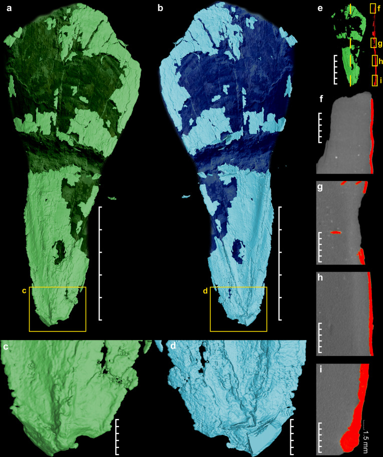
Fig. 2. Micro-CT visualization of the gladius N. hungarica.
a Dorsal view. b Ventral view showing typical traingular median field (scale bars a, b = 5 cm). c detail of the posterior part forming conus, dorsal view. d detail of the posterior part lateral fields expansion, ventral view (scale bars c, d = 1 cm). e Position of lateral micro-CT sections (f–i), scale bar = 5 cm. f–i Lateral gladius sections (red color) documenting rise of thickness towards the apex (conical part), scale bars = 0.5 cm.

