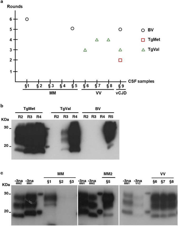
Figure 4.
Detection of PrPTSE in the CSF of CJD patients. CSF samples from 8 sCJD patients (§1–§8) and 1 definite vCJD patients were serially amplified by PMCA. The PrPTSE signal was assessed by means of western blot analysis after proteinase K digestion using 9A2 antibody. For each sample, 20 µL of the product were loaded onto the gel. R refers to the number of rounds. M indicates the typical molecular mass of PrPres in the range of 20–30 kDa. (a): Representation of the results according to CSF sample genotypes and number of rounds; (b): vCJD amplification in TgMet, TgVal and BV substrates; (c): sCJD amplification: MM subtypes in BV substrate; VV subtype in TgVal substrate. − 3na MM2/MM1/VV2 refer to non-amplified material (no PMCA) obtained from the 10−3 dilution (w/v) of the initial infectious brain samples from MM2, MM1 and VV2 subtypes, respectively.

