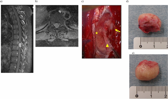
Figure 1.
Resection of meningioma at T8/9 level in a 44-year-old woman. T1-weighted magnetic resonance image (MRI) with gadolinium enhancement. (a) Sagittal and (b) axial views. We measured the maximum tumor length on MRI. (c) Resection of meningioma with inner dural layer (triangle), outer dural layer (arrow), and tumor (star). Excised specimen showing (d) dural side and (e) ventral side. We confirmed attachment of inner dural layer to the tumor.

