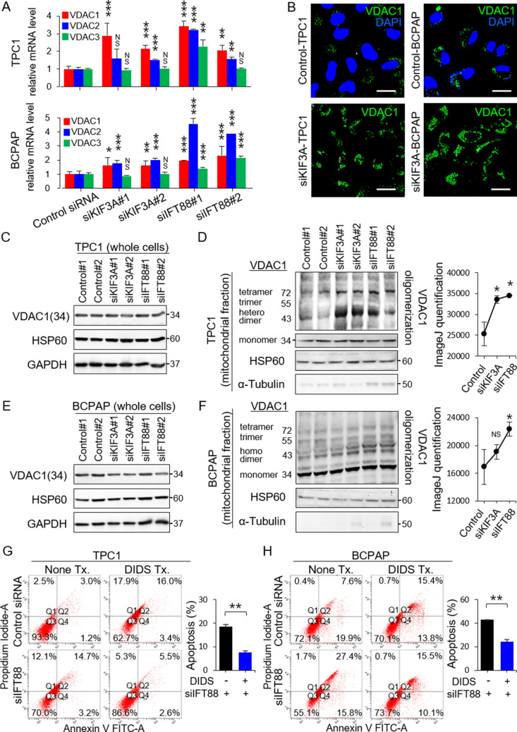
Figure 3.
Loss-of-function of primary cilia in thyroid cancer cell lines upregulates the mitochondria-dependent apoptosis pathway. (A) Expression of VDAC1, VDAC2, and VDAC3 mRNA in human thyroid cancer cells (TPC1 and BCPAP), with or without ciliary loss. **P < 0.01, ***P < 0.001, NS; not significant. (B) Comparison of immunofluorescence staining of VDAC1 between negative control siRNA-transfected cells and KIF3A-deficient TPC1 or BCPAP cells. Scale bar, 10 μm. (C) Western blot analysis of VDAC1 and HSP60 (a mitochondrial volume marker) expression in whole-cell lysates of KIF3A-KD or IFT88-KD TPC1 compared with that in negative control siRNA-transfected cells. (D) Analysis of the oligomeric status of VDAC1 in the mitochondrial fractions of KIF3A-KD and IFT88-KD TPC1 cells compared with negative control siRNA-transfected cells. A line graph was generated from the ImageJ data using arbitrary area units. *P < 0.05. (E) Western blot analysis of VDAC1 and HSP60 expression in whole-cell lysates of KIF3A-KD and IFT88-KD BCPAP cells compared with negative control siRNA-transfected cells. (F) Analysis of the oligomeric status of VDAC1 in the mitochondrial fraction of KIF3A-KD and IFT88-KD BCPAP cells compared with negative control siRNA-transfected cells. A line graph was generated from the ImageJ data using arbitrary area units. *P < 0.05, NS; not significant. (G, H) Flow cytometry analysis of apoptosis in IFT88-deficient TPC1/BCPAP cells treated with DIDS. The levels of apoptosis in IFT88-deficient TPC1 or BCPAP cells treated with DIDS were markedly less than those in untreated IFT88-KD cells. Bar graphs show average percentages of apoptotic cells (Q2 + Q4). **P < 0.01.

