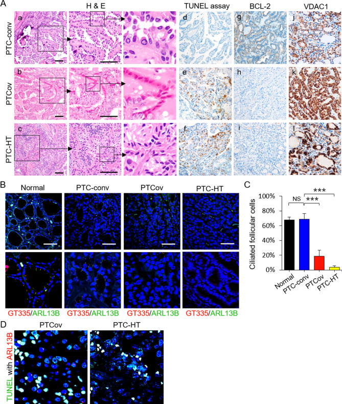
Figure 4.
The proportion of apoptotic cancer cells increases in PTCs showing ciliary loss. (A) Upon histological examination after H & E staining, apoptotic cancer cells appeared dark, with an eosinophilic cytoplasm and dense purple nuclear chromatin fragments (insert). TUNEL analysis revealed significantly more apoptotic cancer cells in PTCov and PTC-HT tissues than in PTC-conv tissue (d, e, and f). BCL-2 expression levels were lower in PTCov and PTC-HT tissues than in PTC-conv tissue (g, h, and i). Immunoexpression of VDAC1 was higher in PTCov and PTC-HT than in PTC-conv (j, k, and l). Scale bar, 10 μm. (B, C) Immunofluorescence images show labeling of primary cilia in normal human thyroid follicles and PTCs by antibodies specific for GT335 and ARL13B. Fewer primary cilia were detected in oncocytic cancer cells in PTCov and PTC-HT tissue than in normal thyroid follicles or conventional PTC tissue. Scale bar, 10 μm. Abbreviations: PTC-conv, conventional papillary thyroid carcinoma; PTCov, oncocytic variant of PTC; PTC-HT, PTC with Hashimoto’s thyroiditis background. ***P < 0.001, NS; not significant. (D) TUNEL with ARL13B double immunofluorescence staining revealed that TUNEL-positive cancer cells (green) have no primary cilia (stained by ARL13B; red).

