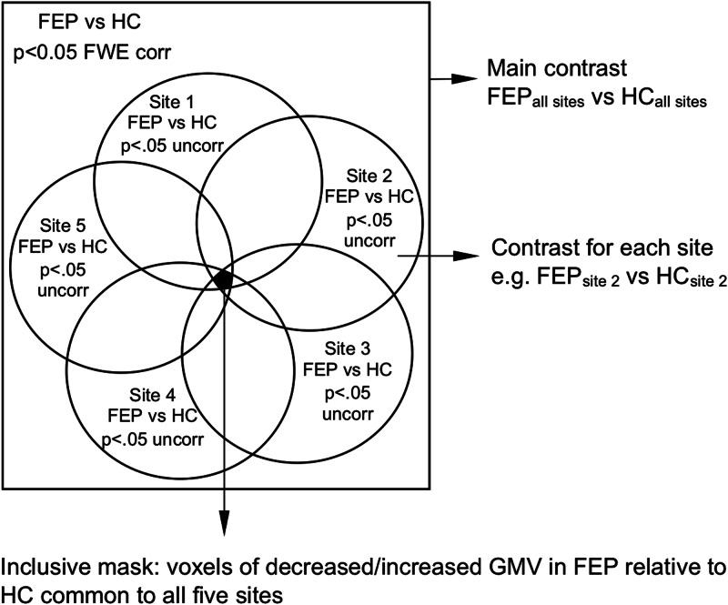
Fig. 1.
Inclusive masking procedure used to identify neuroanatomical abnormalities in FEP relative to HC consistent across all five sites. Left: an overall contrast with all FEP against all HC (p < 0.05 FWE corrected) was combined with five site-level contrasts (p < 0.05 uncorrected); this allowed us to identify only the voxels that survived both types of contrasts (intersection of all contrasts in black).

