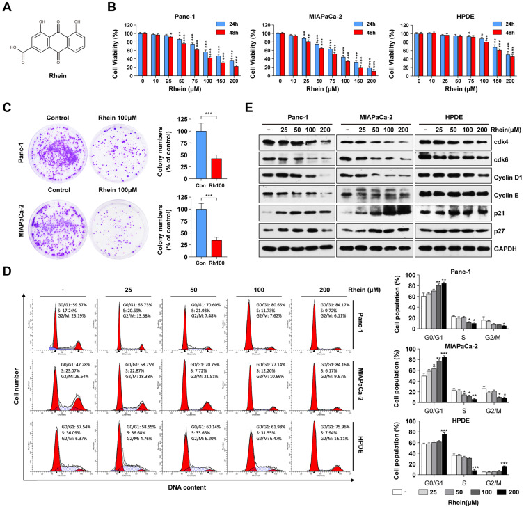
Figure 1.
Rhein inhibits PC cell growth by inducing G1 phase cell cycle arrest. (A) The chemical structure of Rhein. (B) Panc-1, MIAPaCa-2 and HPDE cells were treated with 0-200 μM of Rhein for 24 or 48 h. CCK-8 assays were performed to determine the cell viability. *p < 0.05; **p < 0.01; ***p < 0.001. (C) Panc-1 and MIAPaCa-2 cells were cultured for 14 days to form colonies after treated with 100 μM Rhein or DMSO for 24 h. Representative images are shown. Each bar represents means± SD from three independent experiments. ***p < 0.001. (D) Cell cycle distribution were analyzed in Panc-1, MIAPaCa-2 and HPDE cells after 0-200 μM Rhein treatment. Representative results are shown in the left panel. Statistical comparisons were performed. Each bar represents means ± SD from three independent experiments. *p < 0.05; **p < 0.01; ***p < 0.001. (E) Western blot analysis of protein levels of cdk4, cdk6, cyclinD1, cyclinE, p21 and p27 in Panc-1, MIAPaCa-2 and HPDE cells after indicated concentration of Rhein treatment for 24 h. GAPDH was used as a loading control. Representative results of two independent experiments are shown.

