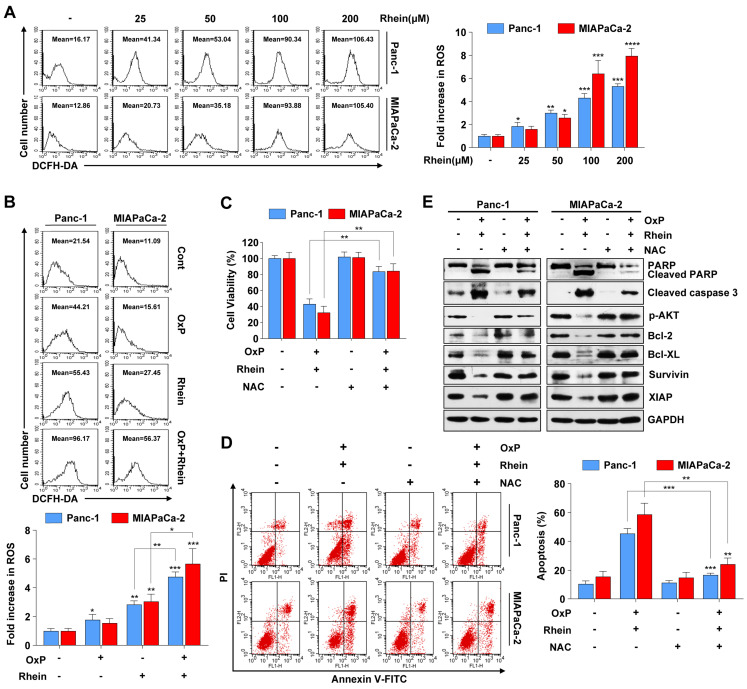
Figure 5.
ROS generation is critically involved in apoptosis induced by the combination of oxaliplatin and Rhein. (A) Panc-1 and MIAPaCa-2 cells were treated with 0-200 μM Rhein for 12 h and incubated with 10 μM DCFH-DA for flow cytometry analysis. Representative results are shown in the left panel. Each bar represents means ± SD from three independent experiments. *p < 0.05; **p < 0.01; ***p < 0.001. (B) Panc-1 and MIAPaCa-2 cells were treated with oxaliplatin (25 μM) or Rhein (50 μM) alone or in combination for 12 h and incubated with 10 μM DCFH-DA for flow cytometry analysis. Each bar represents means ± SD from three independent experiments. *p < 0.05; **p < 0.01; ***p < 0.001. (C) Panc-1 and MIAPaCa-2 cells were pretreated or not with NAC (5 mM) for 1 h, followed by combined treatment of oxaliplatin (25 μM) and Rhein (50 μM). Cell viability was measured by CCK-8 assay. Each bar represents means ± SD from three independent experiments. **p < 0.01. (D) Panc-1 and MIAPaCa-2 cells were treated as described in (C) and apoptosis rates were determined by Annexin V/PI double staining. Each bar represents means ± SD from three independent experiments. **p < 0.01; ***p < 0.001. (E) Western blot analysis of PARP, cleaved-caspase-3, phospho-AKT and Bcl-2 family proteins in Panc-1 and MIAPaCa-2 cells treated as described in (C). GAPDH was used as loading control.

