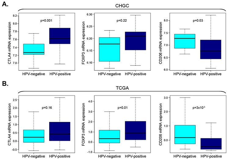
Figure 4. Differences in the tumor microenvironment in HPV-positive and HPV-negative SCCHN tumors.
(A) Box plot analysis of CTLA4, FOXP3 and CD206 mRNA expression in HPV-positive versus HPV-negative TCIP-H SCCHN tumors of the CHGC. CTLA4 mRNA is significantly higher in HPV-positive TCIP-H SCCHN tumors compared to HPV-negative tumors (Mann-Whitney, p=0.001). FOXP3 mRNA is higher in HPV-positive tumors but did not reach significance in this cohort (Mann-Whitney, p=0.22). CD206 mRNA is significantly higher in HPV-negative TCIP-H SCCHN tumors compared to HPV-positive tumors (Mann-Whitney, p=0.03). (B) Box plot analysis of CTLA4, FOXP3 and CD206 mRNA expression in HPV-positive versus HPV-negative TCIP-H SCCHN tumors of the TCGA. CTLA mRNA is higher HPV-positive tumors but did not reach statistical significance in this cohort (Mann-Whitney, p=0.16). FOXP3 mRNA is significantly higher in HPV-positive TCIP-H SCCHN tumors compared to HPV-negative tumors (Mann-Whitney, p=0.03). CD206 mRNA is significantly higher in HPV-negative TCIP-H SCCHN tumors compared to HPV-positive tumors (Mann-Whitney, p=3×10⁻⁵).

