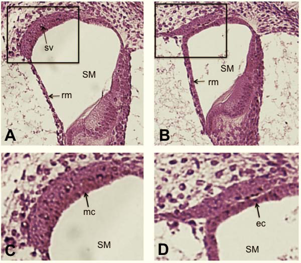
Figure 3. Failure of marginal cell maturation in Tbx1nmf219 mutant mice.

Transverse sections through the cochlear ducts of a +/+ control mouse (A) and a Tbx1nmf219 mutant mouse (B) at P3, stained with H&E. The boxed region of A encloses the stria vascularis and is enlarged in C, and the similarly positioned boxed region of B is enlarged in D. The +/+ control mouse (A) has a normally developed a stria vascularis (sv), but a mature stria vascularis does not form in Tbx1nmf219 mutant mice (B). The arrow in C points to the layer of mature marginal cells (mc) in the stria vascularis of the +/+ control mouse, which have interdigitated with the overlying intermediate cells. The arrow in D points to the layer of precursor epithelial cells (ec) lining the scala media (SM) in the Tbx1nmf219 mutant mouse, which have not matured into marginal cells and do not interdigitate with intermediate cells. For orientation Reissner’s membrane (rm) is marked in A and B.
