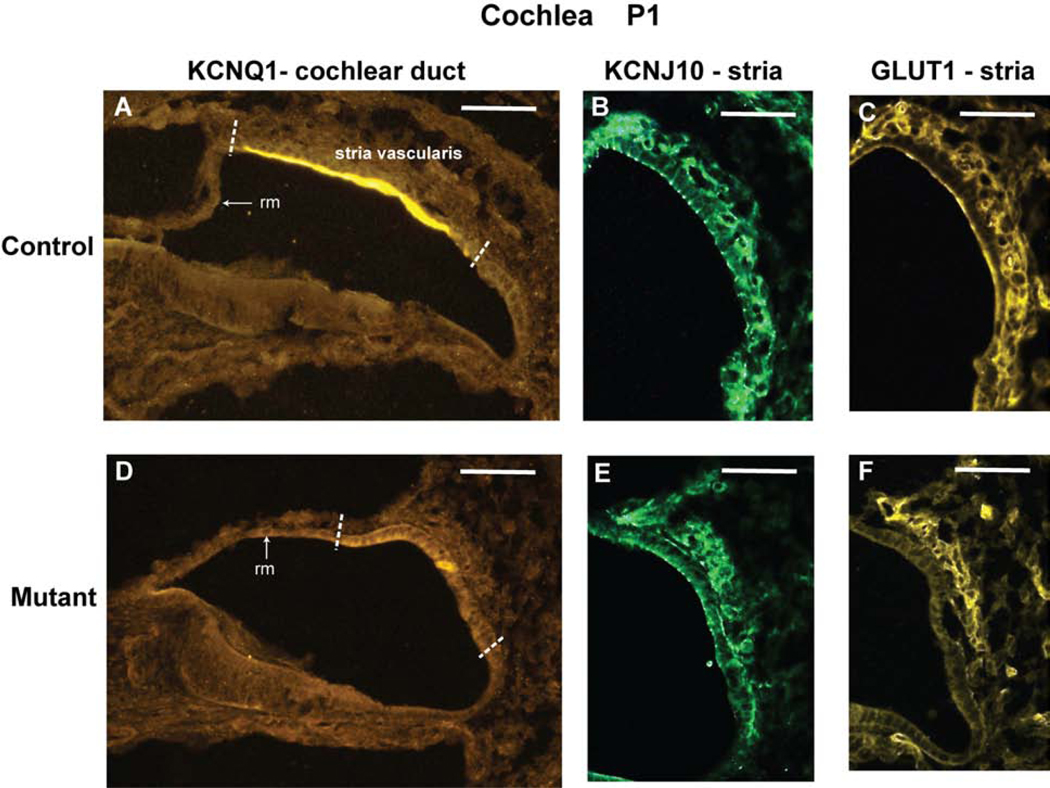
Figure 6. KCNQ1, KCNJ10, and GLUT1 protein expression in cochleae from Tbx1+/nmf219 (control) and Tbx1nmf219/nmf219 (mutant) mice.
The expression patterns of immunolabeled marker proteins for cells of the stria vascularis are shown on transverse sections through the cochlear duct of newborn (P1) mice. The white dashed lines in panels A and D demarcate the approximate location of the developing stria vascularis in the lateral wall of the cochlear duct. Only this region of the cochlear duct is shown in panels B, C, E, and F. KCNQ1 is expressed in marginal cells of the stria vascularis in control mice (A), but its expression was not detected in mutants (D). KCNJ10 is expressed in intermediate cells of the stria vascularis in control mice (B), but its expression is reduced in mutants (E). GLUT1 is expressed in basal cells and intraepithelial capillaries of the stria vascularis in control mice (C), but its expression is reduced in mutants (F). The expression of KCNJ10 (E) and GLUT1 (F) in the cochleae of mutant mice appears stronger in the strial region nearest Reissner’s membrane (rm). The images in B,C and E,F are from different inner ear sections. Scale bars, 50 micrometers.

