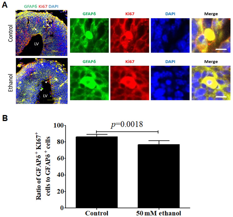
Figure 6.
Effect of PEE on the proliferative activities in GFAPδ+ radial glial cells. (A) In embryonic day 14.5 cortices, co-labeling for GFAPδ and Ki67 demonstrated that GFAPδ+ cells were proliferating RGCs in the VZ, SVZ, IZ or CP. PEE significantly influenced the distribution of Ki67+ cells. In the ethanol cortex, radial glial cells were less abundant for GFAPδ+Ki67+ cells. The arrows indicate examples of GFAPδ+Ki67+ proliferative cells located in the control and ethanol-treated cortex. Arrowheads show the single labeling cells by GFAPδ or Ki67 in the ethanol-treated embryonic cortex. Immunostainings for representative GFAPδ+Ki67+ proliferative cells in control and ethanol groups are showed in the right magnification images. (B) Quantification of the ratio of GFAPδ+Ki67+ positive cells to total GFAPδ+ cells in the cortical layers. Scale bar, 100 µm (Left image) and 10 µm (right magnification images). The P-value was obtained from an unpaired Student's t-test. PEE, prenatal ethanol exposure; GFAP, glial fibrillary acidic protein; SP, subcortical plate; CP, cortical plate; IZ, intermediate zone; VZ, ventricular zone; SVZ, subventricular zone; LV, lateral ventricle.

