Figure 2.
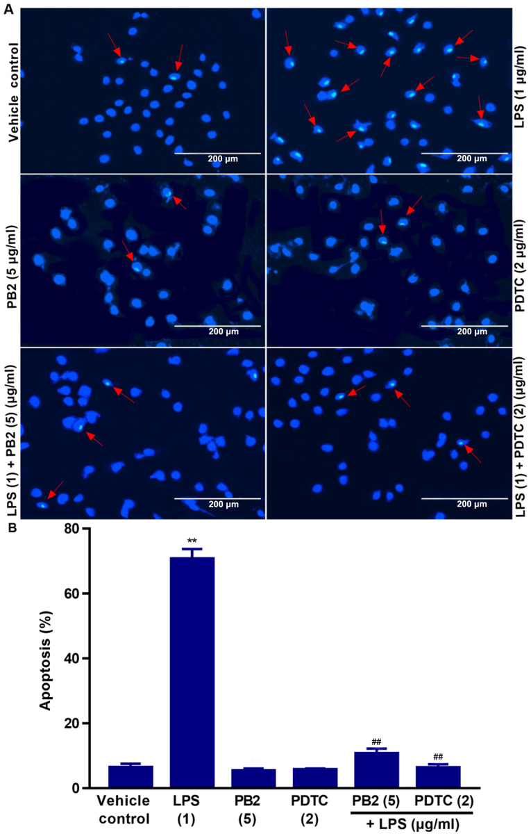
Effects of PB2 and PDTC on LPS-induced HUVEC apoptosis. Cells were treated with serum-free medium alone for 24 h in the vehicle control group; cells were treated with serum-free medium for 12 h followed by LPS (1 µg/ml) for 12 h in the LPS group; cells were treated with serum-free medium for 12 h followed by PB2 (5 µg/ml) for 12 h in the PB2 group; cells were treated with serum-free medium for 12 h followed by PDTC (2 µg/ml) for 12 h in the PDTC group; cells were treated with PB2 (5 µg/ml) for 12 h followed by LPS (1 µg/ml) for 12 h in the LPS + PB2 group; cells were treated with PDTC (2 µg/ml) for 12 h followed by LPS (1 µg/ml) for 12 h in the LPS + PDTC group. (A) Representative images of apoptotic cells identified by Hoechst staining via fluorescence microscopy (magnification, ×400). Red arrows indicate apoptotic cells. The vehicle control group displayed the typical features of HUVECs with the appearance of normal blue fluorescence in the cell nuclei. In LPS-treated HUVECs, apoptotic cells with condensation of nuclear chromatin and fragmentation, which was indicated by white staining, were observed. Pretreatment with PB2 or PDTC prior to LPS treatment markedly reduced the number of apoptotic cells compared with the LPS group. However, PB2 or PDTC treatment alone displayed no obvious effect on HUVEC apoptosis compared with the vehicle control group. (B) Quantification of the percentage of apoptotic cells. Data are presented as the mean ± SD of at least three independent experiments run in triplicate (n=3). Data were analysed using one-way ANOVA followed by Tukey's post hoc test. **P<0.01 vs. vehicle control; ##P<0.01 vs. LPS. PB2, procyanidin B2; PDTC, pyrrolidinedithiocarbamate ammonium; LPS, lipopolysaccharide; HUVEC, human umbilical vein endothelial cell.

