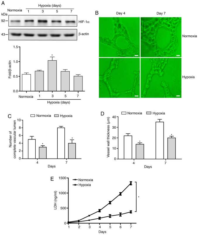
Figure 1.
Sustained hypoxia leads to waveform expression levels of HIF-1α protein and increases the proportion of abnormal blood vessels. (A) Levels of HIF-1α protein in HUVECs cultured under hypoxic conditions for 7 days were examined via western blotting. (B) Angiogenic potential was examined using simulated angiogenesis experiments in HUVECs on days 4 and 7 of exposure to hypoxia (scale bar, 20 µm). (C) The number of complete vascular lumens and (D) vessel wall thickness were measured. (E) LDH content was measured via ELISA following HUVEC exposure to hypoxia for 7 days. Data are presented as the mean ± standard deviation. n=3. *P<0.05 vs. normoxia. HIF, hypoxia inducible factor; HUVEC, human umbilical vein endothelial cells; LDH, lactate dehydrogenase.

