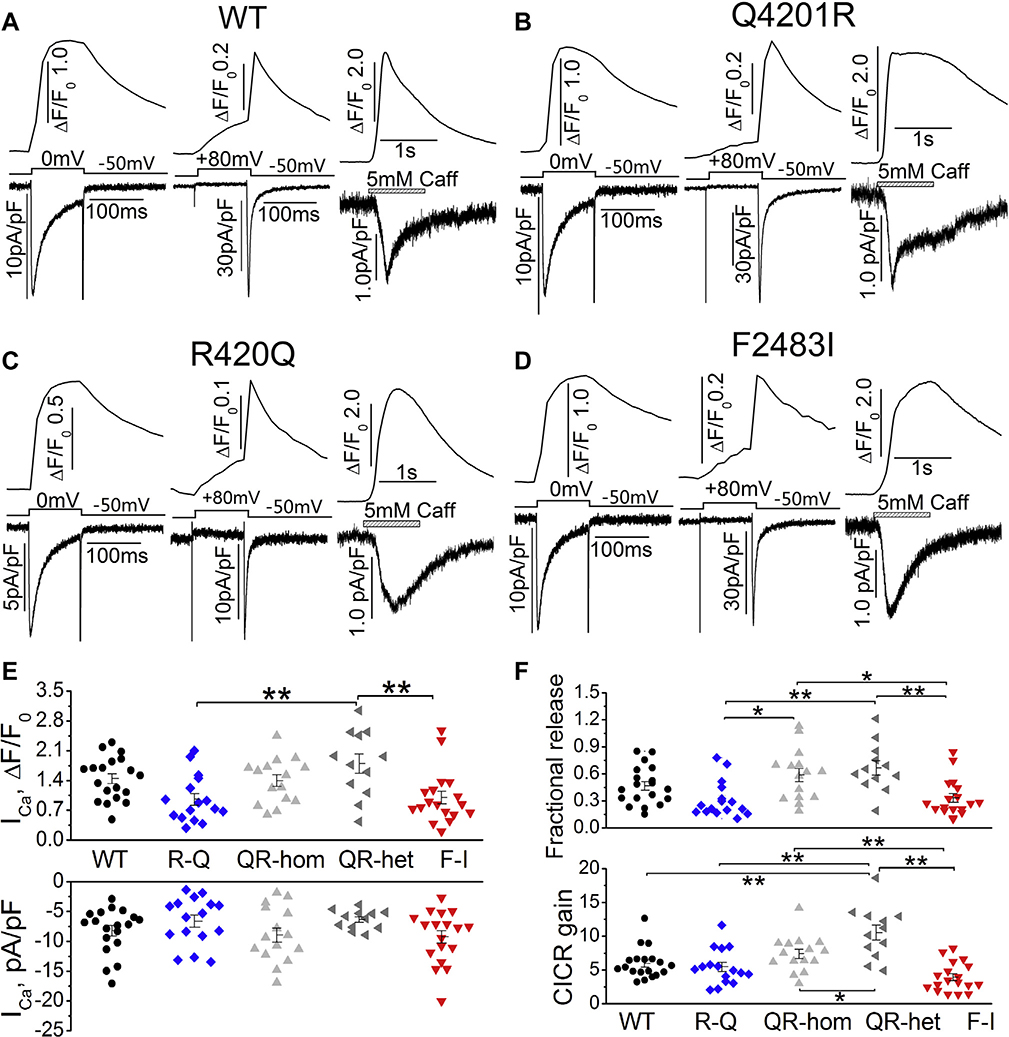
Figure 5.
L-type Ca2+ current(ICa), fractional Ca2+ release, and calcium-induced calcium release (CICR) gain. A–D: Representative traces of ICa and ICa-induced Ca2+ release (left panels), ICa tail currents and their induced Ca2+ release (middle panels), and caffeine-induced Ca2+ release and INCX (right panels) from WT and 3 homozygous mutant hiPSC-CMs. E: Quantified ICa density (bottom panel) and ICa-induced Ca2+ release (top panel) in WT and mutant cells. F: Fractional Ca2+ release (top panel) and CICR gain (bottom panel) in each group. *P < .05, **P < .01 by 1-way analysis of variance.

