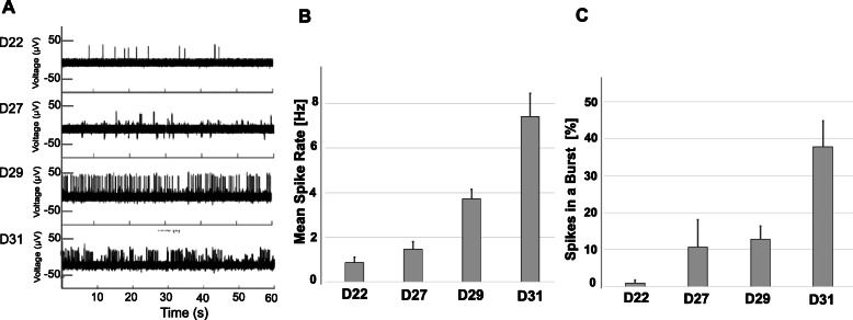
Fig. 2.
Electrophysiological analysis of (MNs). a Time-dependent increase of spontaneous action potential (AP) spikes generated by MNs and recorded from a representative electrode (one of 144) on a multi-electrode (MEA) chip. MN were plated on the MEA chip at 18 days post-chemical induction of iPSC (D18) and electrophysiological activity was recorded at the indicated time points of MN maturation (D22-D31) for 1 min. The frequency of AP spikes (b) and the percentage of spikes in the burst (c) dramatically increased within the neuronal networks over time. Shown are the mean and standard error from 12 electrodes at each sampling day post iPSC induction

