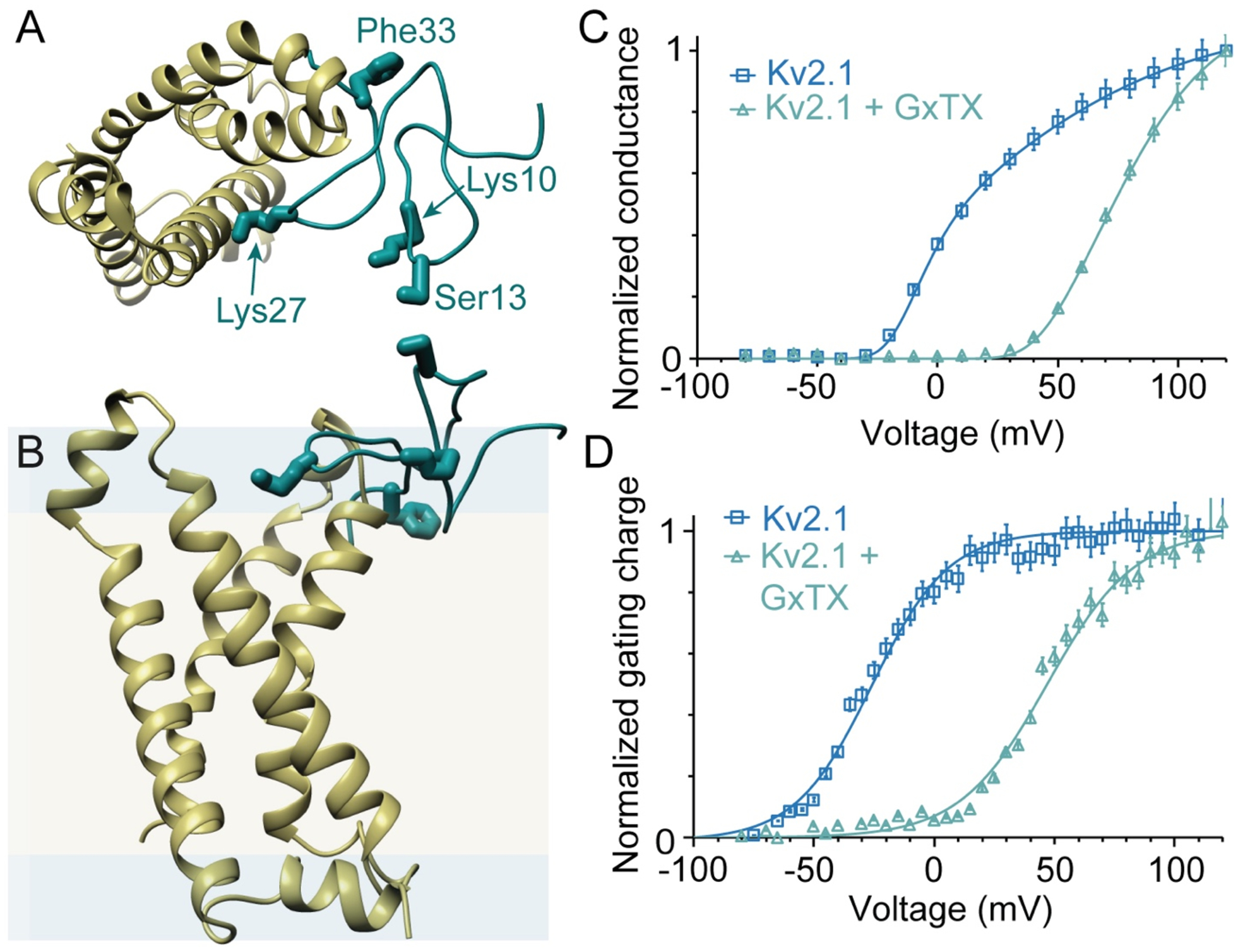
Figure 1.

Structural models and activity of GxTX with Kv channels. (A, B) Top and side views of a model of GxTX (green) bound to a Kv2.1 voltage sensor (gold) in its active state. The GxTx-Kv2.1 VSD model was generated from a Rosetta-based model of the Kv2.1 VSD20 and X-ray structure of the ProTx-II-hNav1.7-NavAb complex.32 The 4 positions of propargylglycine (Pra) substitution and fluorophore conjugation are highlighted. Membrane is shown as hydrophobic interior (yellow) and polar interface (blue), with dimensions calculated using the RosettaMembrane energy function.33 (C) Normalized conductance, G/Gmax, of Kv2.1 with and without 100 nM GxTX, in Kv2.1-transfected CHO cells. (D) Normalized gating charge, Qoff, with and without 1 μM GxTX in Kv2.1-transfected CHO cells. Gating currents measured from Qoff at −140 mV and reported as mean ± standard error of the mean (SEM). Solid lines in (C) and (D) are two-state Boltzmann functions. Data replotted from ref22.
