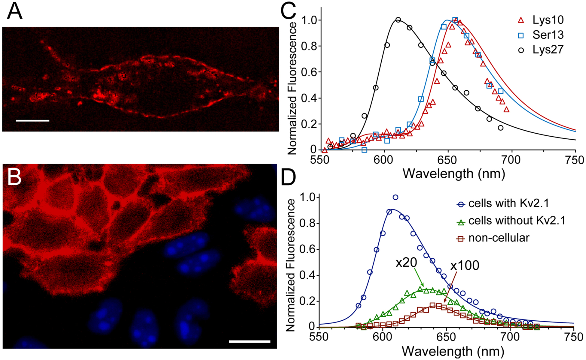
Figure 3.

Fluorescence of JP-GxTX conjugates on live cells. (A) Compressed z-stack confocal image of a live rat hippocampal neuron stained with 100 nM Lys27Pra(JP) GxTX. The cell is excited at 561 nm and its emission isolated around 625 nm. Scalebar is 10 μm. (B) Specificity of JP-GxTX conjugates for Kv2-expressing cells. Confocal image of co-plated CHO cells with or without Kv2.1, stained with 100 nM Lys27Pra(JP) GxTX (red membranes). Only cells without Kv2 channels express nuclear BFP. (C) Spectral imaging of JP-GxTX conjugates on Kv2.1 CHO cells. (D) Emission spectra of Lys27Pra(JP) GxTX imaged from CHO cells with (blue line) or without Kv2.1 (green line, magnified 20x), or from extracellular regions (red line, magnified 100x). Spectra are fit with 2-component pseudo-split Voigt functions, with shape values and root-mean-squared deviations (R2) listed in Table S1.
