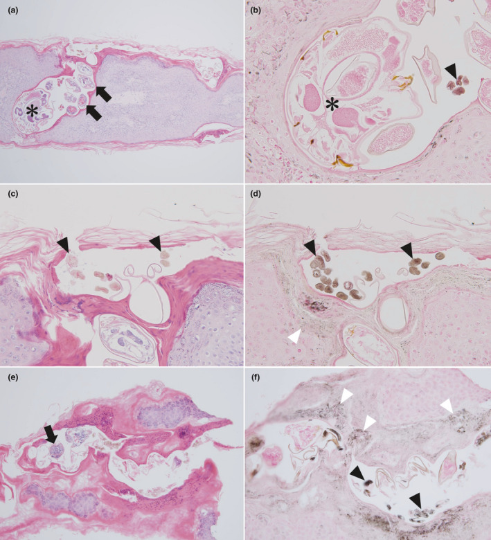
Figure 4.

(a,c,e) Histopathological findings (hematoxylin–eosin, original magnifications: [a] ×100, [c] ×200, [e] ×100) and (b,d,f) Fontana‐Masson staining of scabies burrows showing the gray‐edged line sign ([b] ×400, [d] ×200, [f] ×200). (a–d) A scabies burrow obtained from the right lateral chest in case 15. A mite (asterisk), eggs (black arrows) and feces (black arrowheads) are apparent. Some melanin‐rich keratinocytes are seen in contact with the wall of the scabies burrow (white arrowheads). (e,f) Scabies burrow obtained from the back in case 26.
