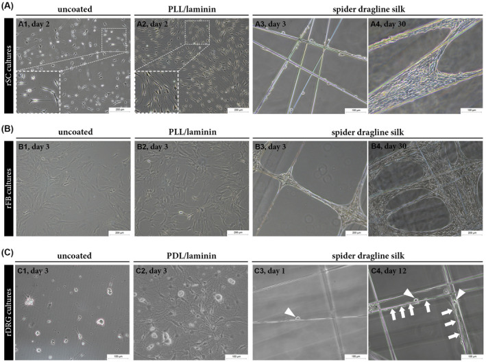
FIGURE 3.

Comparison of the growth of rSCs, rFBs and rDRG neurons on different substrates. A‐C, Representative phase contrast images of rSCs, rFBs, and rDRG neurons cultured on different substrates showing successful adhesion and growth of all cell types on spider dragline silk fibers. A, rSCs cultured on uncoated (A1) and PLL/laminin coated (A2) dishes at day 2 as well as on silk fibers at day 3 (A3) and day 30 (A4). B, rFBs cultured on uncoated (B1) and PLL/laminin (B2) coated dishes at day 4 as well as on silk fibers at day 3 (B3) and day 30 (B4). C, rDRG neurons cultured on uncoated (C1) and PDL/laminin (C2) coated dishes at day 3 as well as on spider silk fibers at day 1 (C3) and day 12 (C4); arrowheads indicate rDRG cell bodies; arrows indicate elongated cells along the silk fibers where rDRGs are present
