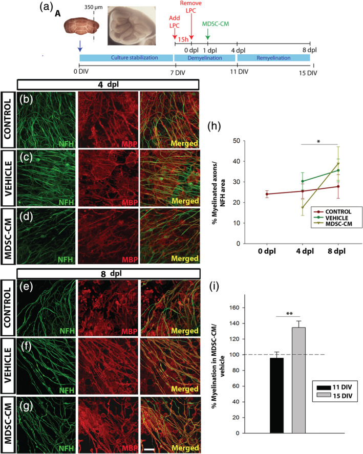
FIGURE 5.

Myeloid‐derived suppressor cell (MDSC) conditioned medium promotes remyelination ex vivo. (a) Experimental procedure for the organotypic culture of cerebellar slices. (b–g): Representative images of the lysolecithin (LPC)‐lesioned slices at 4 (b–d) and 8 dpl (e–g), showing co‐labeling of myelin basic protein (MBP, red) and axons (NFH, green) in the control (b; e), vehicle (c; f), and MDSC conditioned medium (MDSC‐CM) conditions (d; g). (h) Graph showing the remyelinationin culture of the LPC‐lesioned tissue (NFH/MBP co‐labeling relative to the NFH area). (i) Graph showing the increase in myelination (measured as in h) provoked by MDSC‐CM, and compared to the vehicle in nonlesioned slices. Scale bar represents 25 μm in (b–d) and (e–g). The statistical analysis was carried out using a Student's t test (for paired samples) and/or one‐way analysis of variance (ANOVA) (for multicomparison); tests are represented as: *p < .05 and **p < .01
