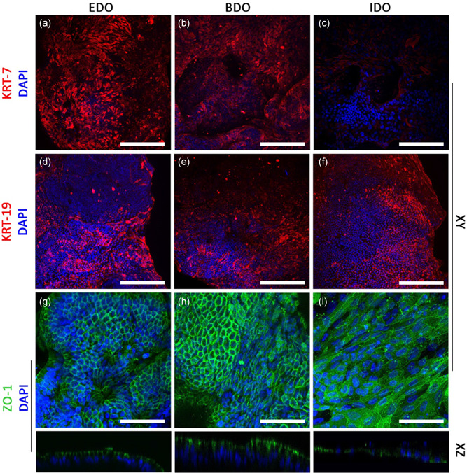
Figure 4.

Organoids derived from EBD tissue or bile samples can be used to repopulate the apical surface of ductal ECM. (a–c) EDO and BDO express cytokeratin‐7 (KRT‐7), whereas IDO showed lower expression of KRT‐7. (d–f) cytokeratin‐19 (KRT‐19) expression was similar between EDO, BDO, and IDO. (k–m) Zone Occludens‐1 (ZO‐1) expression showed that EDO and BDO had a “honey comb”‐like phenotype. ZO1 was located on the luminal side (XZ plane), whereas nuclei were located at the basolateral side, indicating cholangiocyte‐like polarization of the cells. IDO recellularized samples had a flattened phenotype and no polarization of ZO‐1 was detected. Scale bars: (a–f) = 200 µm and (g–i) = 100 µm. BDO, bile‐derived organoids; ECM, extracellular matrix; EDO, extrahepatic bile duct‐derived organoids [Color figure can be viewed at wileyonlinelibrary.com]
