Figure 6.

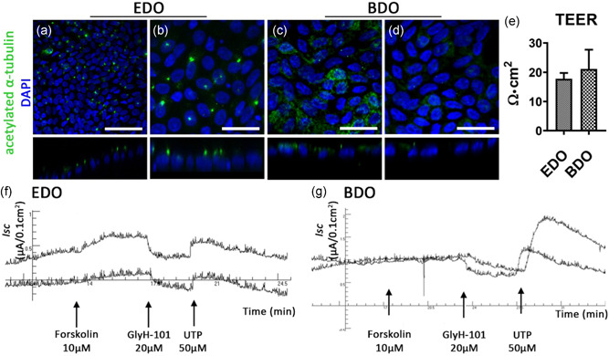
Functional bile duct constructs can be created in vitro using EDO or BDO. (a–d) Acetylated α‐tubulin staining shows the presence of cilia in EDO and BDO recellularized samples. Scale bars: (a) 100 µm, (b–d): 50 µm. (e) TEER measurements show an increase in resistance compared to decellularized ECM (N = 3 measurements per sample). (f) and (g): Ussing chamber experiments showing short circuit current (I sc) changes upon consecutive addition of Forskolin (a cAMP agonist), GlyH‐101 (CFTR inhibitor), and UTP (Ca2+ agonist and CaCC activator) of EDO recellularized ECM (f, N = 2) and BDO recellularized ECM (g, N = 2). After addition of Forskolin a small response in EDO samples was recorded, but not in the BDO samples. GlyH‐101 successfully blocked CFTR‐channel activity in both samples, as a decrease I sc was recorded. Addition of 50 µM UTP showed an increase in current for both samples indicating the presence of CaCC. BDO, bile‐derived organoids; CaCC, calcium activated chloride channels; ECM, extracellular matrix; EDO, extrahepatic bile duct‐derived organoids; TEER, trans epithelial electrical resistance [Color figure can be viewed at wileyonlinelibrary.com]
