. 2021 Feb 19;129(2):027005. doi: 10.1289/EHP7297

Figure 6.

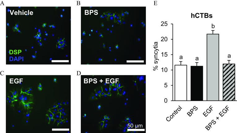
Figures 6A, 6B, 6C, and 6D are a set of syncytialization images of human cytotrophoblasts, and Figure 6E is a bar graph. In Figures 6A, 6B, 6C, and 6D, images of human cytotrophoblasts showing the effects of bisphenol S with 50 micrometers are presented. Vehicle is exposed to 0.1 percent Dimethyl sulfoxide (Figure 6A), bisphenol S is exposed to 200 nanograms per milliliter (Figure 6B), epidermal growth factor is exposed to 10 nanograms per milliliter (Figure 6C), and bisphenol S plus epidermal growth factor is exposed to 200 nanograms per milliliter bisphenol S plus 10 nanograms per milliliter epidermal growth factor (Figure 6D). Nuclei were stained with 4′,6-diamidino-2-phenylindole. The cells were exposed for 96 hours. In Figure 6E, the bar graph titled human cytotrophoblast plots percentage syncytia, ranging from 0 to 25 in increments of 5 (y-axis) across control, bisphenol S, epidermal growth factor, and bisphenol S plus epidermal growth factor (x-axis).

Effect of BPS exposure on human cytotrophoblast (hCTB) syncytialization. Representative images of hCTBs immunostained against desmoplakin (DSP; green) following a 96-h exposure to (A) vehicle (0.1% DMSO), (B) BPS (200 ng/mL), (C) EGF (10 ng/mL), or (D) BPS+EGF. Nuclei were stained with DAPI (blue). (E) The percentage of syncytia [percentage of cells forming a syncytia (3 nuclei)] is represented as mean±SEM. n=6 primary hCTB cell cultures/group. A generalized linear model was used to compare treatments. Different letters denote statistical differences among treatment groups at p<0.05. Note: BPS, bisphenol S; DAPI, 4′,6-diamidino-2-phenylindole; DMSO, dimethyl sulfoxide; EGF, epidermal growth factor; SEM, standard error of the mean.