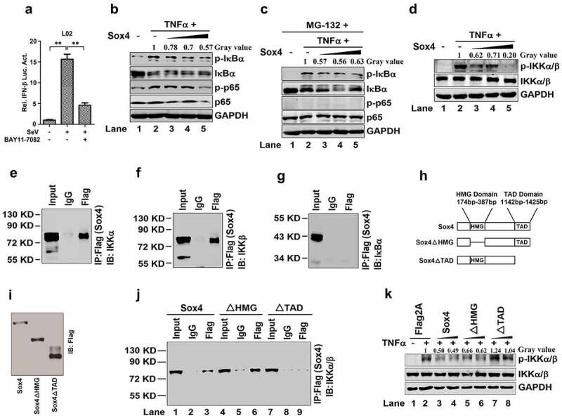
Figure 3.

Sox4 inhibits NF-kB activity through interacting with IKKα/α (a) L02 cells were transfected with pIFN-β-Luc for 24 h, treated with BAY11-7082 (a specific inhibitor of NF-kB) for 9 h, and then infected with SeV for 12 h. Luciferase activities were measured using a TD-20/20 luminometer and normalized to the control. The results are presented as means ± SDs (n = 3). **P < 0.01. (b and c) L02 cells were transfected with pFlag-Sox4 or pFlag2A and treated with TNFα (b) or treated with MG-132 and then treated with or without TNFα (c). The p-IkBα, IkBα, p-p65, p65, and GAPDH proteins expressed in the cells were detected by Western blot analyses. (d) L02 cells were transfected with pFlag-Sox4 or pFlag2A and treated with TNFα. The p-IKKα/α, IKKα/α, and GAPDH proteins expressed in the cells were detected by Western blot analyses. (e–g) HEK293T cells were transfected with pFlag-Sox4. Co-IP assays for the transfected cells were performed using antibody to Flag, and the precipitates were analyzed using antibody to IKKα (e), using antibody to IKKβ (f), or using antibody to IκBα (g). (h) Diagrams of the wild-type Sox4 protein and its two mutants, Sox4∆HMG and Sox4∆TAD. In Sox4∆HMG, the HMG domain of Sox4 was deleted, whereas in Sox4∆TAD, the TAD domain of Sox4 was deleted. (i) HEK293T cells were transfected with pFlag-Sox4, pFlag-Sox4∆HMG, or pFlag-Sox4∆TAD for 48 h. The cells were collected and lysed in Western bolt lyses buffer. Sox4, Sox4∆HMG, and Sox4TAD proteins were analyzed by Western blot using antibodies specific to FLAG. (j) HEK293T cells were transfected with pFlag-Sox4, pFlag-Sox4ΔHMG, and pFlag-Sox4ΔTAD, respectively. Co-IP assays were conducted using antibody to Flag, and the precipitates were analyzed using antibody to IKKα/β. (k) L02 cells were transfected with pFlag2A, pFlag-Sox4, pFlag-Sox4ΔHMG, and pFlag-Sox4ΔTAD, respectively, and treated with TNFα. The p-IKKα/α, IKKα/α, and GAPDH proteins expressed in the cells were detected were detected by Western blot analyses
