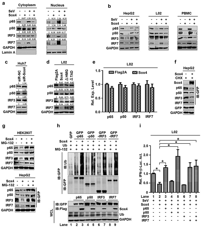
Figure 4.

Sox4 downregulates NF-kB and IRF3/7 by facilitating protein degradation. (a) L02 cells were transfected with pFlag-Sox4 and infected with SeV. Proteins expressed in the cytoplasm (left) or nucleus (right) were detected by Western blot analyses. (b–f) HepG2 cells, L02 cells, and PBMCs were transfected with pFlag-Sox4 and infected with SeV (b). Huh7 cells were transfected with siR-Sox4 (c). L02 cells were transfected with pFlag-Sox4, pFlag-Sox4ΔHMG, or pFlag-Sox4ΔTAD (d). L02 cells were transfected with pFlag2A or pFlag-Sox4. Total mRNA extracts were prepared from the cells. The p65, p50, IRF3, and IRF7 mRNAs expressed in the cells were determined by RT-PCR using the corresponding primers (e). HepG2 cells were co-transfected with pFlag-Sox4 and pGFP-p65, pGFP-p50, pGFP-IRF3, or pGFP-IRF7, and treated with cycloheximide (f). 293 T (left) and HepG2 cells (right) were co-transfected with pFlag-Sox4 and pGFP-p65, pGFP-p50, pGFP-IRF3, or pGFP-IRF7, and treated with MG-132 (g). p-p65, p65, IRF3, IRF7, Sox4, GFP, and GAPDH proteins were detected by Western blot analyses. (h) L02 cells were co-transfected with pMYC-Ub and pFlag-Sox4, pGFP-p65, pGFP-p50, pGFP-IRF3, or pGFP-IRF7, and treated with MG-132. Proteins in cell lysates were detected by Western blot analyses, proteins in supernatant were detected by IP assays or by Western blot analyses. (i) L02 cells were co-transfected with pIFN-β-Luc and pFlag-Sox4, pGFP-p65, pGFP-p50, pGFP-IRF3, or pGFP-IRF7, and infected with SeV. Luciferase activities were measured using a TD-20/20 luminometer. The results are shown as means ± SD (n = 3). *P < 0.05
