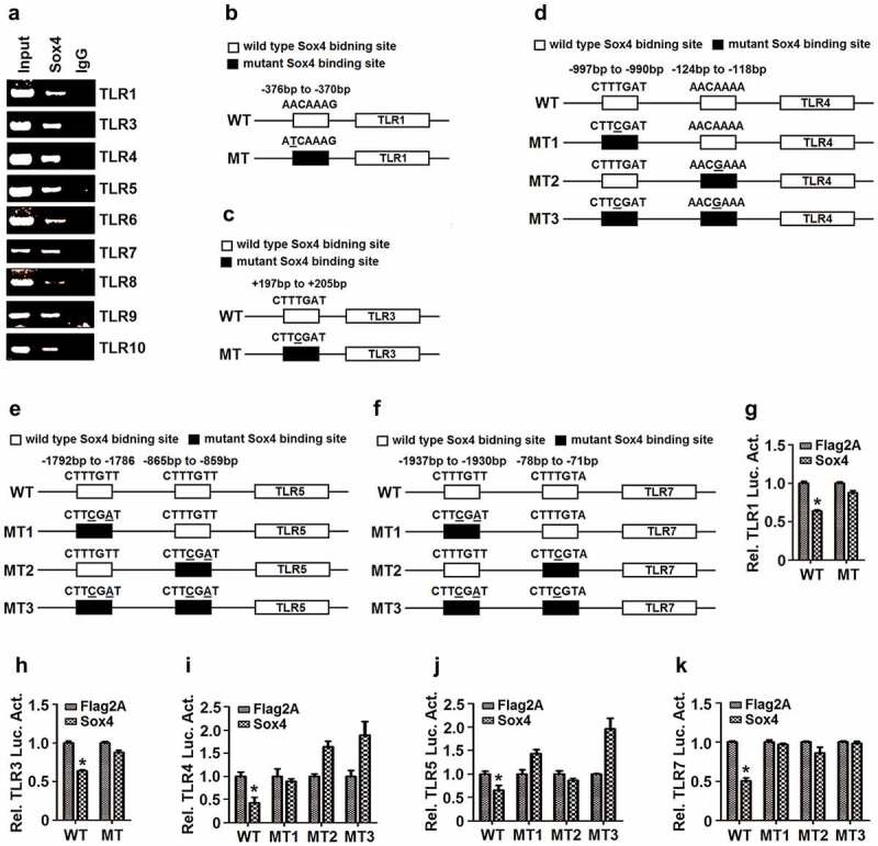
Figure 7.

Sox4 represses TLRs expression by binding to the promoters. (a) L02 cells were transfected with pFlag-Sox4. The cell extracts were prepared for IP analyses using antibody to Flag, and the precipitated DNA were analyzed by PCR using ChIP primers for TLR1, TLR3, TLR4, TLR5, TLR6, TLR7, TLR8, TLR9, or TLR10. (b–f) Diagrams of the WT-TLR1 and MT-TLR1 promoters (b); the WT-TLR3 and MT-TLR3 promoters (c); the WT-TLR4, MT1-TLR4, MT2-TLR4, and MT3-TLR4 promoters (d); the WT-TLR5, MT1-TLR5, MT2-TLR5, and MT3-TLR5 promoters (e); and the WT-TLR7, MT1-TLR7, MT2-TLR7, and MT3-TLR7 promoters (f). □ The wild-type of Sox4 binding site on TLR promoter; ■ The mutant Sox4 binding sites on TLR promoter, in which the mutated nucleotides are underlined. (g–k) L02 cells were co-transfected with pFlag-Sox4 along with pWT-TRL1-Luc or pMT-TRL1-Luc (g); with pWT-TRL3-Luc or pMT-TRL3-Luc (h); with pFlag-Sox4 and pWT-TRL4-Luc, pMT1-TLR4-Luc, pMT2-TLR4-Luc, or pMT3-TLR4-Luc (i); with pWT-TRL5-Luc, pMT1-TLR5-Luc, pMT2-TLR5-Luc, or pMT3-TLR5-Luc (j); and with pWT-TRL7-Luc, pMT1-TLR7-Luc, pMT2-TLR7-Luc, or pMT3-TLR7-Luc (k). Luciferase activities in the cell extracts were measured using a TD-20/20 luminometer. The results are shown as means ± SD (n= 3). **P< 0.05
