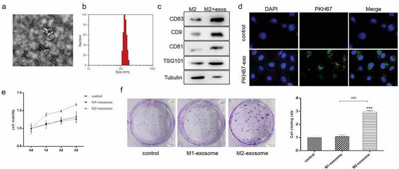
Figure 3.

Exosomes derived from M2 macrophage promoted the proliferation and tumorigenesis of GC cells. (A). Scanning electron microscopy of exosomes isolated from conditioned medium of M2 macrophage. (B). Nanoparticle tracking analysis (NTA) of M2 macrophage-exosomes isolated by ultracentrifugation. (C). Western blot analysis of exosome markers. Tubulin was used as internal control. (D). Immunofluorescence analysis of the internalization of PKH67-labeled M2 macrophage-exosome (green) by HS-746 T cells. (E & F). HS-746 T were treated with M1 macrophage-exosomes, M2 macrophage-exosomes (100 ug/ml) or no-treatment. After 24 h, MTT was used to test cell viability of HS-746 T (E), and colony formation assay was used to detect cell proliferation (F). ***p < 0.001 vs control. ###p < 0.001 vs M1 macrophage-exosomes group. Data are displayed as mean ± SEM
