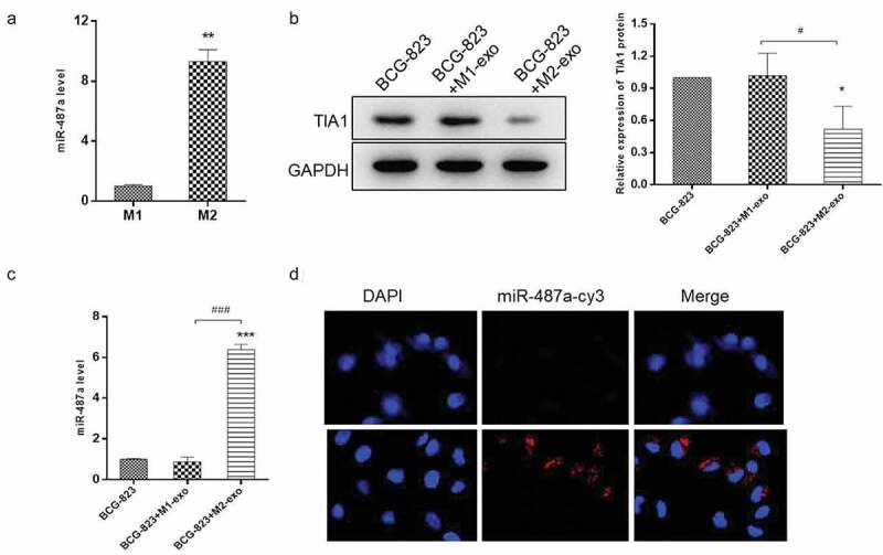
Figure 4.

miR-487a is highly expressed in M2 macrophage-exosomes and can be transferred to GC cells via exosomes. (A). qRT-PCR were performed to detect the expression level of miR-487a in M1 and M2 macrophage-exosomes. (B & C). HS-746 T cells were treated with M1 macrophage-exosomes, M2 macrophage-exosomes or no-treatment. After 24 h, the expression level of miR-487a were detected by qRT-PCR (B), and the protein level of TIA1 were analyzed by western blot (C). GAPDH were used as the internal control. (D). Immunofluorescence analysis of the internalization of Cy3-labaled-miR-487a mimic (red) by HS-746 T cells. *p < 0.05, ***p < 0.001 vs control. #p < 0.05, ###p < 0.001 vs M1 macrophage-exosomes group. Data are displayed as mean ± SEM
