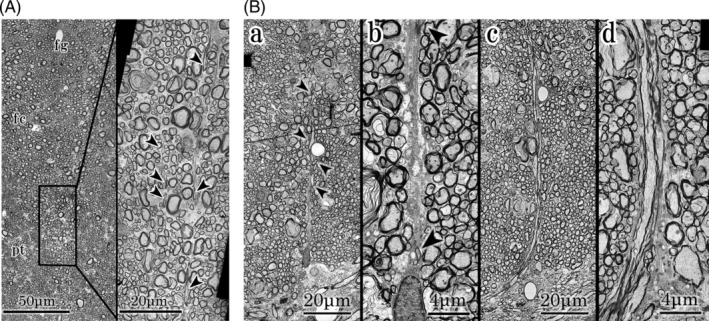
FIGURE 3.

A: Higher magnification of ventral part of fasciculi gracilis and median part of fasciculi cuneatus. Left;Ventral part of fasciculi gracilis and median part of fasciculi cuneatus. Axons of right and left fasciculi gracilis and cuneatus fuse at the median part. fg; ventral part of fasciculi gracilis, fc; central part of fasciculus cuneatus, pt; pyramidal tract. Right; Higher magnification of the central part of fasciculi cuneatus. Several tangential axons (arrow heads) are noted at the median part of the fasciculi cuneatus. B: Higher magnification of ventral part of fasciculi cuneatus and gray commissure. a,b; specimens of Figure 1. From a triangular glial protrusion, a short lineal figure of glial tissue extends dorsally. Tangential axons (arrow heads) are noted in the glial tissue. c,d; contralateral specimens. Three to four axons enter the gray commissure vertically. Bundles of astrocytes extend along the axons from a triangular glial protrusion
