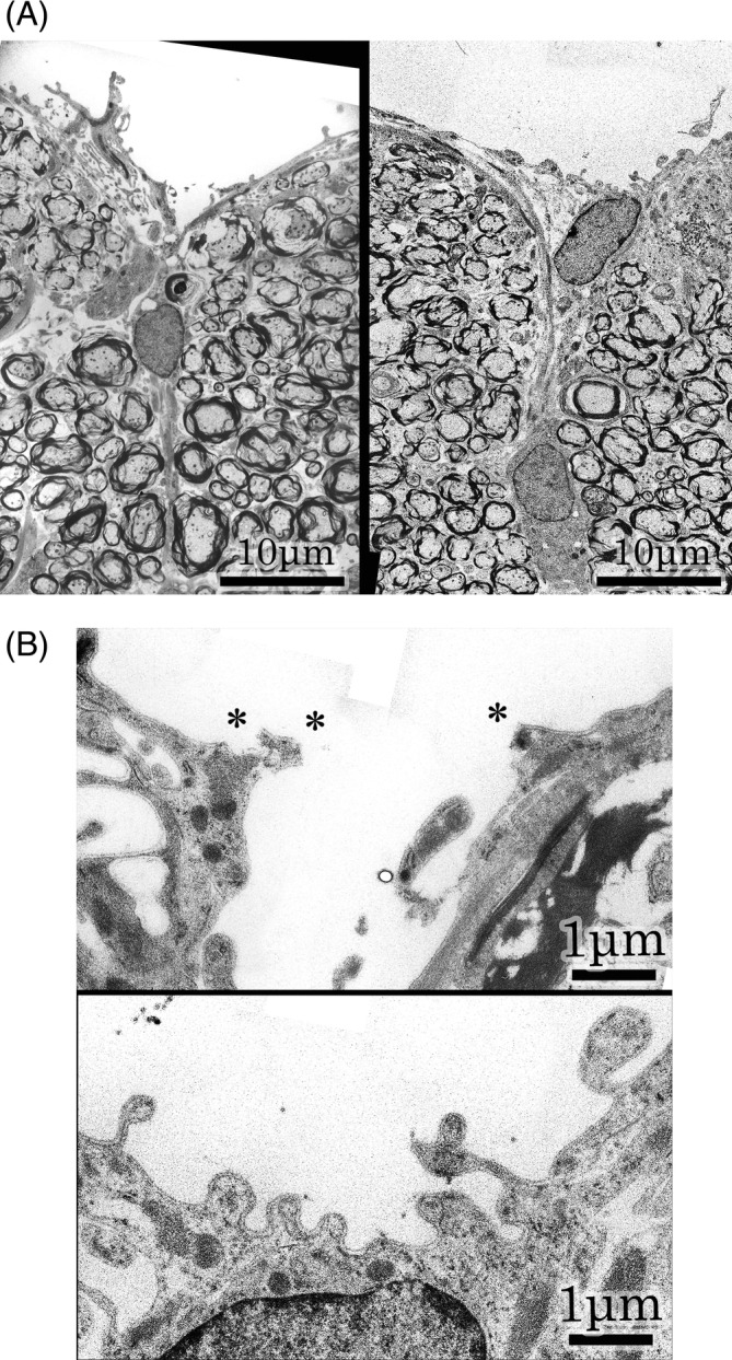
FIGURE 4.

A: Posterior median sulcus. Left ; Magnification of Figure 1 specimen. Posterior median sulcus shows an acute angle. The groove is covered superficially with glial tissues, continuing to the bilateral glial membranes. Right ; Magnification of contralateral specimen. Posterior median sulcus is a shallow depression. A triangular glial tissue is recognized under the posterior median sulcus. Glial membranes are contiguous with a midline lineal figure of glial tissue. B : Higher magnifications of posterior median sulcus. Upper ; Higher magnification of Figure 1 specimen. Basal lamina is partially disrupted at the star marks. The glial tissues split to right and left. Lower ; Higher magnification of contralateral specimen. The posterior median sulcus is lined with basal lamina of astrocytic process
