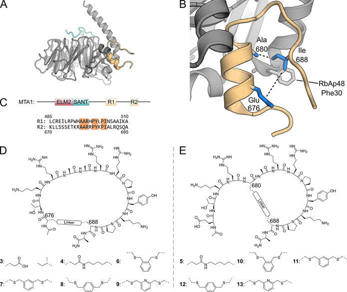
Figure 1.

A) RbAp48 with the MTA1 R2 fragment (residues 670–695, orange) bound to the flank binding site. The FOG‐1 peptide (residues 1–13, cyan) is bound to the top site. Superimposition of PDB files 4pbz and 2xu7. B) Zoom of crystal structure of RbAp48 bound by MTA1 R2 peptide (residues 670–695, PDB: 4pbz). [10] Indicated are the peptide positions used for cyclization (blue side chains). C) MTA1 domain structure and sequences of the MTA1 R1 and R2 binding sites. Identical amino acids in both binding sites are highlighted. D) Structure of cyclic peptides 3, 4, 6–9. E) Structure of cyclic peptides 5, 10–13.
