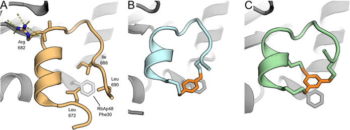
Figure 2.

A) Crystal structure of MTA1 R2 peptide bound to RbAp48 (PDB: 4pbz). [10] Shown are the three amino acids forming the hydrophobic cluster around Phe 30 of RbAp48 and the hydrogen bonding network of Arg 682. B) Peptide 8 bound to RbAp48. The xylene linker is highlighted in orange. (PDB: 6ZRC) C) Peptide 33 bound to RbAp48. The mesitylene linker is highlighted in orange. (PDB: 6ZRD).
