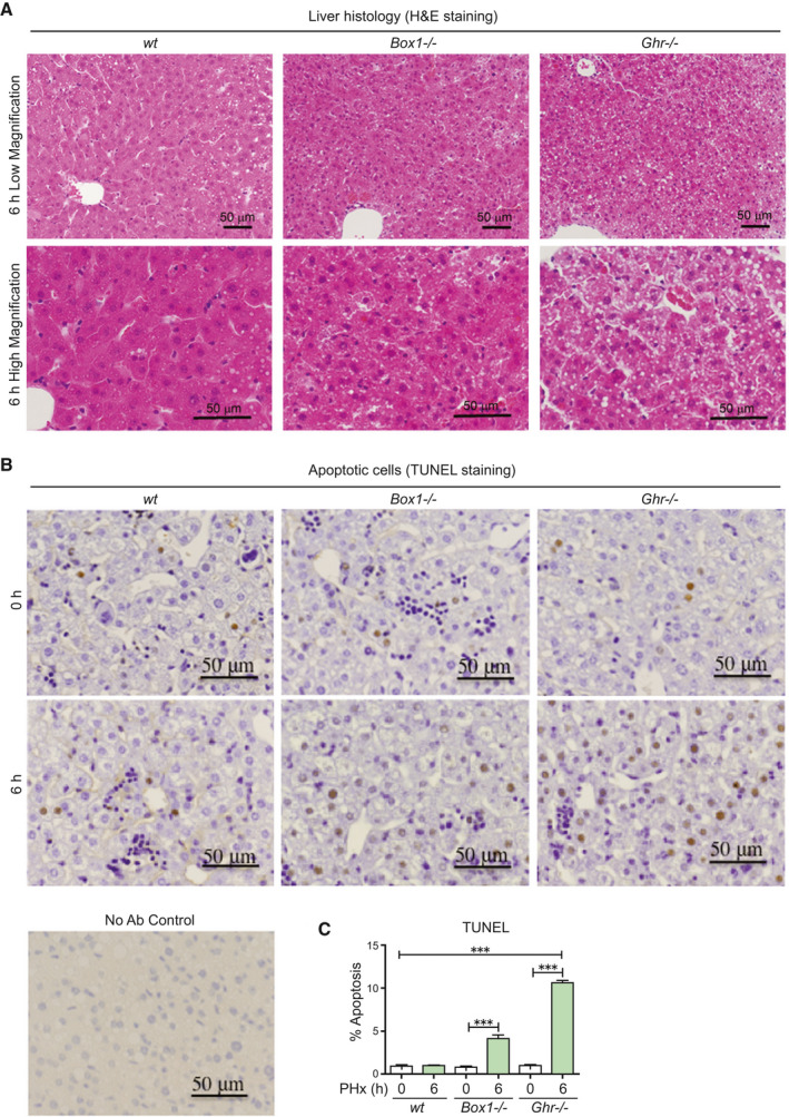
FIG. 2.

The livers of Ghr −/− mice 6 hours following PHx show lipid droplets, ballooning, and increased apoptosis compared with wt and Box −/− mice. (A) Liver histology from Box1 −/− and Ghr −/− mice compared with wt 6 hours after PHx, showing lipid droplets and ballooning in Ghr −/− liver. Lower panel shows high magnification images. (B) TUNEL staining in livers at 0 hours and 6 hours after PHx, showing increased apoptosis in Box1 −/− mice compared with wt, but particularly in Ghr −/− mice compared with wt. Staining without primary antibody is shown (No Ab Control). (C) TUNEL quantification in livers at 0 hours and 6 hours after PHx. Six to nine sections were analyzed for each mouse at 0 hours and 6 hours following PHx, with n = 4‐11 mice per group. Abbreviation: H&E, hematoxylin and eosin.
