
FIG. 5.
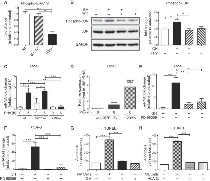
GH activates JUN and up‐regulates H2‐Bl and HLA‐G expression in a SRC‐ERK pathway–dependent manner, protects hepatocytes from NK cell attack, and H2‐Bl expression is induced in livers of wt and Box1 −/− C57BL/6 mice after PHx, but not in Ghr −/− C57BL/6 mice. (A) ERK1/2 activation in Ghr −/− liver 6 hours after PHx is impaired compared with Box1 −/− and wt mice (quantification from western blot, n = 3 for each mouse line). (B) Immunoblot showing suppression of GH‐stimulated JUN phosphorylation by SRC kinase inhibitor PP2 (10 μM) in AML12 hepatocyte cells and its quantification (n = 3 wells per condition). (C) H2‐Bl transcript level does not increase after PHx in Ghr −/− , whereas wt and Box1 −/− show a significant increase (n = 3‐6 per group). (D) Basal expression of H2‐Bl transcript in the 129/Sv mouse strain is high compared with C57BL/6. (E) GH treatment increases H2‐Bl transcript in AML12 mouse hepatocyte cells, and this increase is blocked by the MEK inhibitor PD98059 (20 μM) (n = 3 replicates per group). Inhibition of ERK1/2 is shown in Supporting Fig. S3. (F) GH treatment increases HLA‐G transcript in human hepatocyte cells, and this increase is blocked by the MEK inhibitor PD98059 (20 μM) (n = 3 replicates per group). Quantification of TUNEL staining of human hepatocytes following co‐culture with human NK cells and treatment with either GH (G) or HLA‐G (H) protein. Abbreviations: GAPDH, glyceraldehyde 3‐phosphate dehydrogenase; PP2, pyrazolopyrimidine (PP)2; and rRNA, ribosomal RNA.