Figure 1.

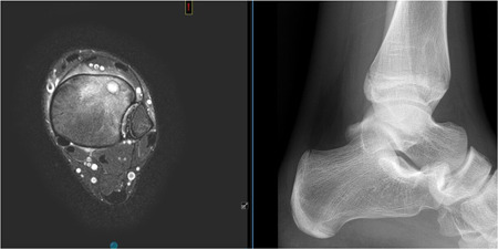
MRI and radiographic view of a Cierny I endomedullary osteomyelitis of the distal tibia (anterolateral). MRI, magnetic resonance imaging [Color figure can be viewed at wileyonlinelibrary.com]

MRI and radiographic view of a Cierny I endomedullary osteomyelitis of the distal tibia (anterolateral). MRI, magnetic resonance imaging [Color figure can be viewed at wileyonlinelibrary.com]